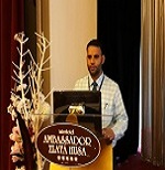
Showkat Hussain Tali
AIMSR, India
Title: Poster 4: Post-operative stridor following repair of tracheoesophageal fistula: A case repor

Biography:
Showkat Hussain Tali is working as Assistant Professor Pediatrics, Adesh University. After obtaining his Bachelor's degree in 2005, he obtained his MD in Pediatric Medicine from University of Kashmir in 2010. In 2013 he joined Department of Neonatology at Surya Children’s Hospital, Mumbai and became Board Certified in Neonatology from the National Board of India in 2016. In the same year, he joined Adesh University as Assistant Professor Pediatrics and In-charge Neonatology. He has more than a dozen publications in national and international journals. He has received Science Talent Search Award from the Govt. of Jammu and Kashmir in 1997 and has been awarded by Help Foundation and Rajiv Gandhi Foundation, India, for excellence in creative writing in 2007. On May 26/2017, he presented a speech at International Congress of Gynecology and Obstetrics, Prague, Czech Republic and has been invited to deliver speech at International Congress of Pediatrics, Taiyuan China (Nov 2017).
Abstract:
A full term, male infant with no significant antenatal and birth history developed severe respiratory distress on day 2 of life. Infant was diagnosed to have H-type of tracheoesopheageal fistula (TEF) and was operated for the same on day 4 of life. Infant was extubated on day 20 of life (difficult extubation) and was put on HHHFNC (heated humidified high flow nasal cannula). Soon after extubation, infant developed severe respiratory distress and stridor. Infant was put back under ventilator support. Flexible laryngoscopy along with bronchoscopy was performed under light sedation. Except for mild subglotic edema, no abnormality was detected. Size 3.5 ET (endotracheal) tube was replaced with a 3 size ET tube and a short course of dexamethasone (0.2 mg/kg/day × 5 days) was administered. After a 10 days period, the infant could be weaned to CPAP (continuous positive airway pressure). However it was not possible to take the infant off the CPAP thereafter. CECT (contrast enhanced computed tomography) was performed and no significant abnormality was detected. Parents were counseled for a tracheostomy but they refused. After one month period, when there was no improvement in clinical condition, laryngoscopy with bronchoscopy was again performed under anesthesia. Tight aryepiglotic folds were detected and aryepiglotic split was performed. Infant responded dramatically to treatment and could be weaned to room air within 3 days of surgery. The anesthesia technique has been found to be superior to awake technique with a sensitivity, specificity, positive predictive value and negative predictive value of 100% each as compared with 93%, 92%, 97%, and 79%, respectively, for awake technique. Most probably, we missed the diagnoses in the first place as we didn’t perform the laryngoscopy under anesthesia or sufficient sedation. It is worth mentioning that laryngoscopy along with bronchoscopy and esophagoscopy was performed under anesthesia during the initial evaluation of TEF before surgery. This makes us strongly believe that the tight aryepiglotic folds were a complication of TEF repair surgery or prolonged intubation rather than a congenital one.